Plen Medical-Goat Ultrasound Machine Center
Your Reliable Goat Ultrasound machine manufacturer in China
The economic benefits of goat farms are directly related to the reproductive characteristics of goats, and goat ultrasound plays a significant role in the diagnosis of goat pregnancy.
Through goat ultrasound examination, the pregnancy of the female goat can be determined.
Breeders/veterinarians can analyze goat ultrasound testing results to scientifically raise pregnant female goats by grouping and independent feeding to improve pregnant female goats’ nutritional management level and increase the lambing rate.
Goat ultrasound is a high-tech method for cross-sectional observation of goats without any damage and irritation.
It has become a beneficial assistant for goats and sheep’s diagnosis activities and a necessary monitoring instrument for scientific research such as live egg collection and embryo transfer.
At this stage, the most commonly used method to check the pregnancy of female goats is to use a goat ultrasound machine.
Goat ultrasound is usually used for goat pregnancy diagnosis, disease diagnosis, estimation of litter size, identification of stillbirth, etc.
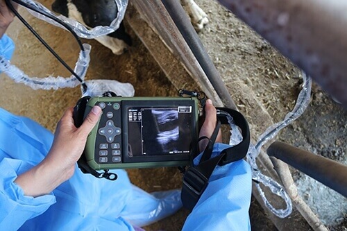
Portable Veterinary Mobile Ultrasound for Goats PM-V1S
Goat ultrasound has the advantages of rapid examination and obvious results.
Compared with the traditional inspection methods in the past, goat ultrasound greatly simplifies the inspection process.
It reduces the inspection cost, which is conducive to the owner/veterinarian quickly finding the problem and adopting the response plan faster, such as rapid grouping.
Goats use a goat ultrasound machine to carry out a goat pregnancy examination, an accurate, fast, and simple imaging technique.
The application of transrectal or transabdominal ultrasound can reach nearly 100% accuracy.
For this reason, most farms have begun to use mobile goat ultrasound machines to test the pregnancy of female goats.
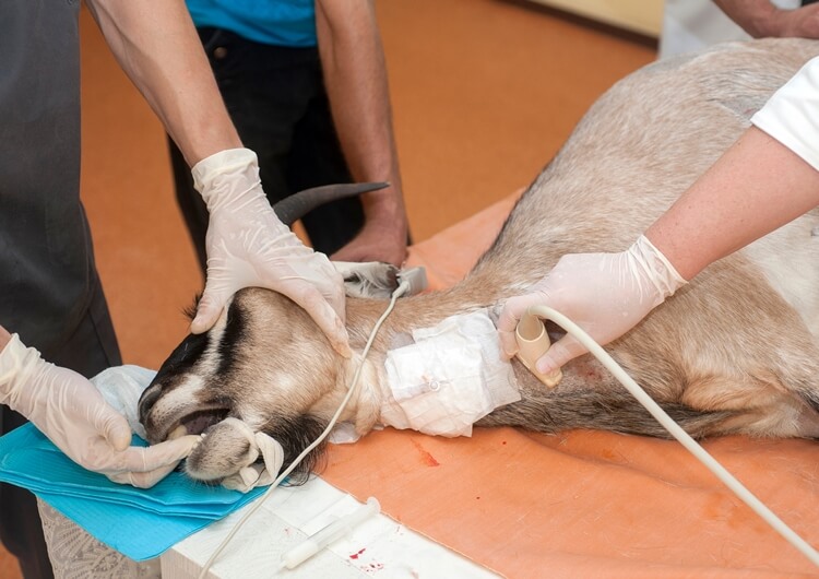
In 2021, the number of consultations for goat farmers has increased significantly.
It can not only measure pregnancy but also estimate gestational age and litter size.
A good goat ultrasound machine can also detect uterine diseases, which helps farmers keep reasonable, save costs and improve efficiency.
The ultrasound diagnosis for goats can also be tested through progesterone and blood. Blood tests are usually called “PAG” tests.
However, blood tests have a high error of 20%, and when determining single births and multiple births, It’s not very accurate either.
At present, the pregnancy test method for goats uses goat ultrasound to check goats, which is cheaper, safer and more accurate.
The goat ultrasonic machine can also detect whether the fetus is still alive so that the fetus is not found to be stillborn when the female goat is giving birth.
Pregnancy testing for extension is generally done by abdominal testing. The best time to detect pregnancy is between 45 and 90 days for female goats.
One of the advantages of goat abdomen detection with goat ultrasound is that it can easily and accurately detect pregnancy, confirm whether the fetus is alive, and the number of pregnant female goats.
1. Doing ultrasound for goats is helpful for farm management, keeping pregnant and non-pregnant goats separately from breeding and management;
2. The pregnancy status can be detected in the abdomen 35 days after mating, and the pregnancy status can be detected in the rectum after 25 days of mating;
3. Doing goat ultrasound can detect multiple births or single. Therefore, single and multiple birth goats can be managed according to nutritional requirements, and feed can be saved.
It is a simple process to do an ultrasound for a female goat. It only requires a portable ultrasound machine and coupling for a goat, and it can be operated by someone nearby.
The pregnancy can be detected 35-40 days after mating and 80-100 days after mating. Is more accurate.
(1) Exploring the abdominal wall of the exploration site.
In the early pregnancy, explore the sparse hair area between the two breasts and the space between the two breasts.

The right abdominal wall can be explored in the middle and late stages of pregnancy. No shearing is required for exploration in the hairless area.
Shearing is required for lateral abdominal wall exploration and rectal exploration.
(2) Detection method
The detection method is basically the same as that of pig ultrasound—the examiner squats on the side of the goat’s body.
Then, after applying the coupling agent on the part or probe, the probe is pressed against the skin and directed towards the entrance of the pelvic cavity to perform a fixed-point fan-shaped scan.
Scan from front to back of the breast, from both sides to the middle of the breast, or from the middle to both sides of the breast.
The fetal sac is not large in the first trimester, and the embryo is tiny. It needs slow scanning and careful examination to detect
(3) The examiner can also squat behind the goat’s buttocks and hold the probe from the middle of the goat’s hind limbs to the breast for scanning.
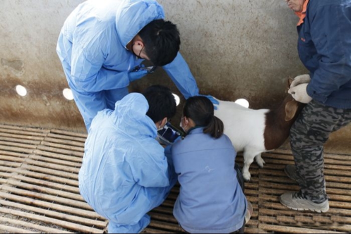
If the breast of the milk mountain goat is too large, or the coat on the side of the abdomen wall is too long, which affects the inspection site, the assistant can lift the hind limb of one side to expose the inspection site, but it is not necessary to cut the hair.
Goat ultrasonic examination of female goats means restraining the female goats generally stands in a natural standing posture.
The assistant can support them and keep quiet, or the assistant can clamp the female goat’s neck with both legs to keep the female goats’ stability or use a simple restraining frame.
Side-lying fixing can slightly advance the diagnosis date and improve the diagnosis accuracy, but it is inconvenient to use in large groups.
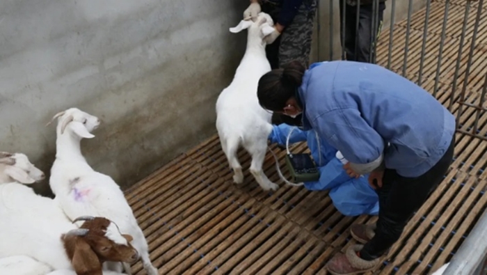
Goat ultrasound exploration of early pregnancy can be performed on the side, supine or standing.
To distinguish between false images, we must recognize several typical goat ultrasound images.
1) Ultrasonic imaging features of female goat’s follicles on goat ultrasound:
Most of them are round in shape, and a few are elliptical and pear-shaped.
From the echo intensity of goat’s B image, due to the internal follicle filled with follicular fluid, the goat was anechoic when scanned by goat ultrasound, and the image was a dark area, which formed a clear contrast with the strong echo (bright) area of the follicular wall and surrounding tissues.
2) Features of goat ultrasound imaging with corpus luteum:
Most corpus luteum tissue is round or oval in shape. Since the ultrasound scan of the corpus luteum tissue is a weak echo, the color of the goat ultrasound image shows that there is no dark color of the follicle.
In addition, the biggest difference between the ovary and the corpus luteum in the goat ultrasound image is the presence of trabecula and blood vessels in the corpus luteum tissue.
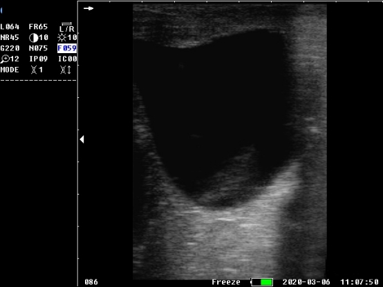
So there are scattered points and bright areas on the imaging, but the follicles do not.
After the inspection, the goats to be inspected are marked and grouped.
1. Do not use the instrument close to generators, dentistry, broadcasting and other equipment. Otherwise, it may interfere with the image and affect the quality of the image.
2. Before using goat ultrasound for the first time, you must carefully read the goat ultrasound machine instruction manual and operate in strict accordance with the requirements.
3. During the operation, do not unplug the probe and protect its surface. Instead, you can rub the coupling agent on the surface of the probe so that the tested animal and the probe have good contact.
4. Practice a more proficient operation process and check quickly.
5. Treat goats gently and not violently grabbing. Otherwise, it may cause problems such as miscarriage of goats.
The goat ultrasound cost is generally about 600-900 US$.
Of course, some better goat ultrasound machines with a high price of more than 1000$.