Plen Medical-Pig Ultrasound Machine Center
Your Reliable Pig Ultrasound machine manufacturer in China
With the application of pig ultrasound monitoring technology and an early determination of non-pregnant pigs, can also early scientific diagnosis of pigs reproductive disorders (abnormal ovarian function or disease, uterine disease, stillbirth, abortion, testes, accessory glands and other diseases of boars).
Take measures such as treatment, elimination or aphrodisiac to improve sow productivity and avoid economic losses.
In addition, the breeding pig farm can accurately measure the backfat thickness of pigs and calculate the area of eye muscles without injury in vivo by pig ultrasound, which greatly improves the scientificity and accuracy of breeding and selection.
The use of pig ultrasound in pig farms helps pig farmers plan reproductive management, optimize artificial insemination programs, reduce reproductive failures, and monitor the physical condition of pigs and improve the overall profitability of the farm.
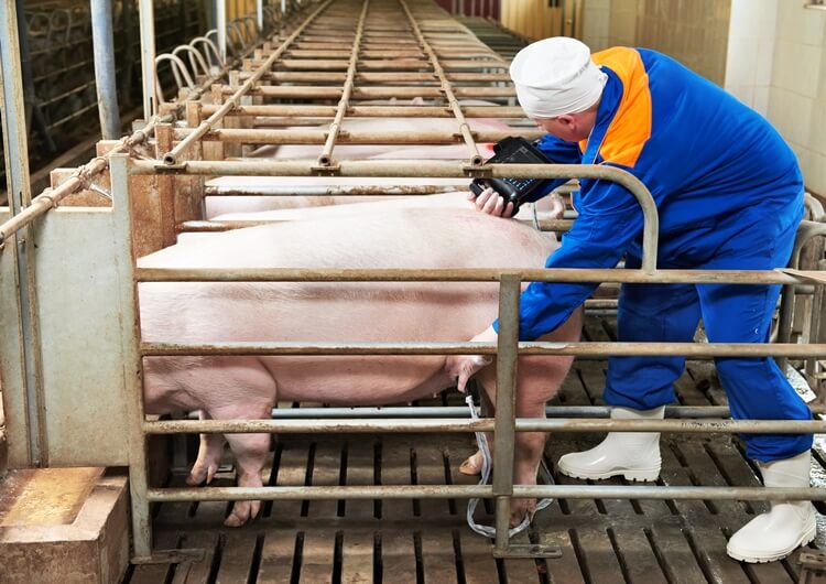
Several common problems in farms can usually be checked strategically with pig ultrasound:
The main purpose of the pig ultrasound examination is pregnancy screening and checking the number of pig litters.
The pregnant pigs can be quickly identified with pig ultrasound machines from the pigs that have never been conceived, and good breeding management can be carried out to reduce the number of non-productive days.
At the same time, the non-pregnant pigs can be eliminated or used as the target of the next cycle for reproduction.
Moreover, the decline in pregnancy rate may be an early sign of farm disease, sometimes even 12 weeks before any clinical symptoms, allowing us to take preventive action earlier.
Ultrasound scans of pregnancy pigs show multiple fluid-filled sacs in the uterus.
The fluid appears black on the pig ultrasound scan, and the uterus is white, so it is easy to identify.
Why is pig ultrasound examination better than other inspection methods?
a. The inspection can be started with a pig ultrasound machine in the early pregnancy of pigs (the inspection can be started from the 20-21 days after insemination, and the scan on the 28th day is more effective because the fluid-filled sacs in the uterus are larger and more obvious, especially in inexperienced people In the case of scanning.
b. This is especially true if it is a free-range pig and the pig moves around. The contact time between the pig and the ultrasonic probe is minimal before the pig runs away. )
C. With a pig ultrasound machine, the pregnancy accuracy in the first-trimester examination is over 97%.
d. In addition to regular pregnancy tests, pig pregnancy failures can also be tested.
For example, by checking the absence of a heartbeat or checking the blood flow signal of the umbilical cord, stillbirth can be detected.
Unfortunately, the blood flow signal of the umbilical cord can only be detected by doppler pig ultrasound.
In addition, pig ultrasound can directly see the death or miscarriage of embryos and confirm the diagnosis.
E. Pig ultrasound examination can be carried out through the skin of the abdomen, instead of going through the rectum like horses or cows.
The best way to check the pig ovary is a transrectal examination with a pig ultrasound machine.
Under pig ultrasound examination, all structures around the ovary and ovary during ovulation can be clearly displayed.
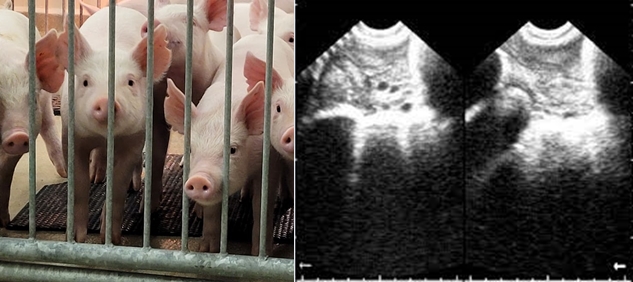
Even after the end of ovulation, the ovaries can be clearly identified based on the presence of corpora hemorrhagic. Therefore:
Based on this, we can estimate the time between weaning and oestrus.
Monitoring method:
Use pig ultrasound 1-2 times a day, from the time that all follicles reach 6mm in diameter until no follicle structure can be seen.
Due to the persistence of ovulation, some follicles will exceed 10mm in diameter, and hemorrhagic bodies from newly ovulated follicles can also be seen.
When pre-ovulation follicles are not detected, it means that ovulation has been completed. This is because the ovaries cannot be seen when the bleeding body is dominant.
Generally speaking, the prepubertal stage of a pig is about 5 months old.
At this time, the uterus is located at the level of the pelvic lymph nodes, at the end of the bladder, away from the abdominal wall, so the effect of transabdominal pig ultrasound is not good.

When the pig is more than 5 months old, the ovaries begin to develop, the uterus begins to enlarge, and the two uterine horns begin to move forward to the level of the bladder.
The uterus is even larger and more forward in pigs at the third and fourth lumbar vertebrae levels.
In pregnant pigs, the uterus can even be located at the level of the last thoracic vertebra, and the uterine horns can touch the abdominal wall.
However, the ovary position is always at the level of the sixth lumbar vertebra on the dorsal side.
Introducing breeding pigs is a critical step for the farm, and introducing them at the wrong time will cause economic losses.
Usually, the detection of ovulation can be determined by measuring the first fever state, but in sexually mature pigs, there are about 7%-20% of negative results.
Therefore, sometimes the first ovulation cannot be monitored by this method.
Pig ultrasound has no concerns in this regard. It is not affected by the degree of sexual maturity of the sow, and its reliability can be as high as 97%.
In immature sows (immature sows), the uterus has a uniform echo structure under pig ultrasound images.
Still, it may be difficult to locate the uterine horns and ovaries, while sexually mature sows can easily locate the uterine horns and ovaries. As a result, the uterine echo looks very uneven.
As the gilts become sexually mature, the uterus begins to develop from prepuberty to puberty.
After the first ovulation of the ovaries, the sows begin to ovulate periodically, and the corpus luteum in the ovaries continues to appear.
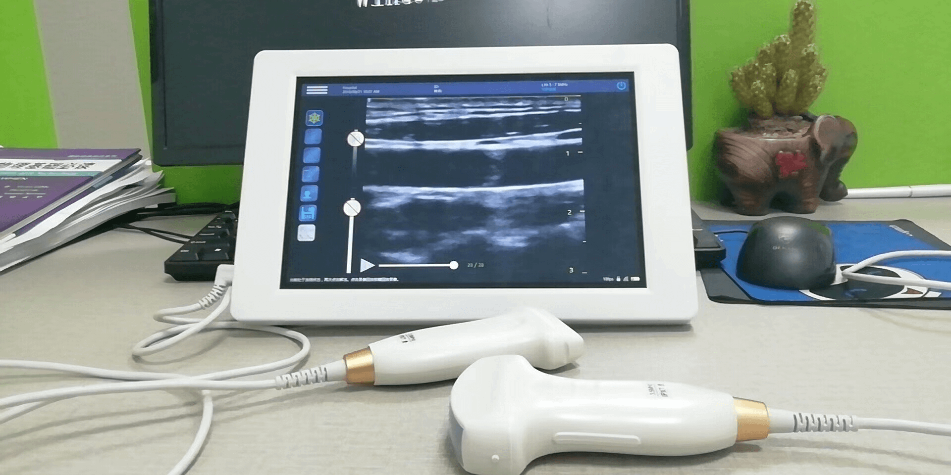
Mobile USB Veterinary Pig Ultrasound Probes
Pig ultrasound can very intuitively see the progress of the uterus and ovaries from prepuberty to puberty.
This is because there are only small vesicles in prepuberty, and in puberty, large vesicles and the corpus luteum that mark the completion of ovulation can be detected.
To date, its accuracy is 100%, which provides precious information for identifying the sexual maturity of sows.
It is almost as reliable to detect the sow’s progress into puberty and detect changes in the size of the uterus as it is to detect changes in the ovaries.
Uterine inspection angle: cross-sectional imaging, and then measure and calculate the cross-sectional area in two dimensions.
Data reference: prepubertal cross-sectional area ≤1cm² (diameter ≤0.9cm), puberty cross-sectional area ≥1.2cm² (diameter ≥1.1cm).
In addition, it is the most accurate to detect the uterus and ovaries at the same time to check the sexual maturity of the sow.
There are many reasons for reproductive failure, but if it is caused by a defect or disease related to the ovary or uterus, then swine ultrasound is directly helpful.
a. Both transabdominal pig ultrasound and transrectal swine ultrasound can detect: ovarian cysts of follicular origin, anovulatory follicles, ovarian cysts of corpus luteum origin, abnormal development of the corpus luteum with trabecular structure (usually> 12mm in diameter) )etc.

b. The only ones that can be diagnosed by pig ultrasound are POD. Transrectal pig ultrasound can calculate the number of cystic structures to diagnose polycystic ovary, one of the most common reproductive diseases in pigs.
This is a type of polycystic ovarian degeneration (POD). The ovary has only a cystic structure, which is fatal to fertility.
The number and size of cysts can be seen intuitively in the ultrasound scan of pigs.
The sows with POD can be quickly removed, reducing the number of sows that cannot reproduce.
c. Inactive ovaries are also one of the reasons for pig reproductive failure. Small follicles characterize pig ultrasound (<6mm), and pigs have a long time of estrus after weaning or no fever within 7 days after weaning. These are often due to high ambient temperature and insufficient metabolism in summer, which leads to "second litter syndrome" in sows. e. Many sows with reproductive failure have problems with uterine infections. Although chronic infections are more common, swine ultrasound machines can confirm and detect acute infections.
Diagnosis method: Acute endometritis can be diagnosed by detecting abnormal flocculent or coagulated fluid in the uterus by ultrasound scanning of pigs, combined with observation of vaginal purulent secretions.
Please note that in the examination of the reproductive system of pigs, except for semen, heat, or pregnancy, the presence of any other fluid is considered pathological.
f. Other diseases, including para ovarian cysts, hematomas or tumors, may cause reproductive failure and be detected by pig ultrasound.
For sows: In addition to the above inspections, it is also important to monitor body conditions (BCS) at different stages, at least after weaning, the first month of pregnancy, and before delivery.
When the sow is too thin, she may have fertility problems or prolific issues.
When the sow is overweight, her legs may have problems, and delivery problems may occur. Unhealthy sows will lead to a high herd replacement rate and cause farm losses.

Before the pigs are sent to slaughter, the back fat and muscles of the pigs can be examined using pig ultrasound. Some companies also measure muscles in the waist and eye area.
It is generally to check whether the IMF (intramuscular fat) or marbling in the muscle is up to the standard.
This helps the farm obtain better quality meat, producing more juicy and delicious pork, getting a better selling price, and creating higher profits.
And many slaughterhouses also use ultrasound in their production lines.
To select high-quality breeding pigs, the farm should carry out an ultrasound examination of the reproductive system of the boars.
This allows 15-20% of poor-quality boars to be screened out to breed truly high-quality piglets.
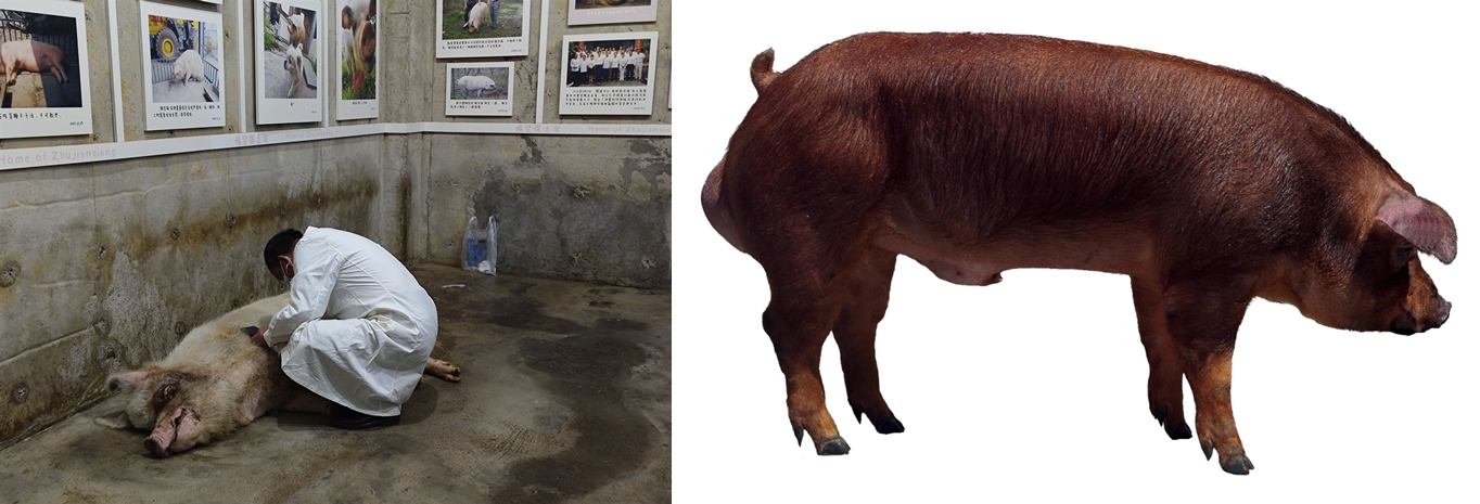
Specific inspection method:
For transrectal swine ultrasound, a 5/7.5 MHz probe is used. However, for best results, a 5/10 MHz linear probe can be used.
Carefully scan the different levels of the testis, the head, body, tail and the three accessory gonads of the epididymis.
The times are constantly developing, and breeding technology is constantly being updated.
Now more and more farmers are accepting new technologies to develop the breeding industry better.
For example, the development of veterinary pig ultrasound machines has given more and more farmers more benefits.
Large-scale intensive pig farms should adhere to routine and institutionalized monitoring.
The time is to monitor once every half month, and the number of days after mating should be 20 to 35 days.
In the past conventional early pregnancy diagnosis methods, for sows that were not estrus after mating in the first estrus and were not pregnant (accounting for about 20% of the number of mating heads).
They generally need to wait until about 40 to 80 days after mating (determined by the level of veterinary technology ) can be more surely diagnosed.
Such sows may cause ineffective feeding for about 20 to 60 days in a breeding cycle.
According to the experimental pig farm statistics, the accuracy of pregnancy diagnosis after using pig ultrasound for early pregnancy monitoring increased by at least 9%.
Every time an empty sow is found, 20-60 days of ineffective feeding can be reduced, and the feeding cost can be saved by 20-60$ (calculated at 1$ per day).
For example, if a pig farm with 1000 large-scale pigs has 200 sows, In case of emptiness, direct economic losses can be reduced by 4000 to 12,000 US$.
Calculated based on the 114-day gestation period of each adult sow, each adult sow produces 2.3 litters per year.
If the sow is empty or suffers from reproductive disorders, it will inevitably affect the number of reproductive sows.
Combined with market conditions, it is invisible Lieutenant will cause greater losses.
The key to the production benefit of large-scale pig breeding lies in balanced production. Balanced production depends on balanced breeding.
Ultrasound monitoring of pigs can accurately grasp the number of pregnant sows. At the same time, pig ultrasound can observe the uterine involution of sows after delivery.
There are data in this regard.
For example, it is reported that if used in production, sows with normal reproductive function can be selected to participate in inbreeding.
The number of healthy sows participating in the breeding can be accurately grasped, and the conception rate during estrus can be increased to ensure balanced production.
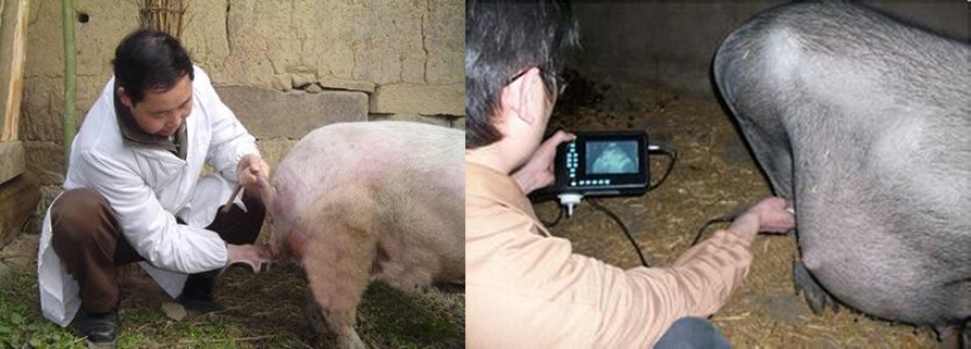
Through pig abdominal ultrasound, only follicles with a diameter greater than 7mm can be detected, so it is impossible to fully monitor all the growing follicles, the structural continuity of the ovaries, and the uterus.
And only about 20-50% of follicles and corpus luteum can be detected by abdominal ultrasound.
Pig transrectal ultrasound can detect follicles with a diameter of about 6mm, most of the corpora lutea and hemorrhages, and the detection is more efficient and comprehensive.
Through pig transabdominal ultrasound, for the monitoring of pregnancy of sows, pregnancy can be seen from the 21st day after insemination.
Pig transrectal ultrasound can be checked 12-16 days in advance.
There is not much difference between pig transabdominal and transrectal ultrasound.
The advantage of transrectal ultrasound is that the structure will be displayed more clearly, but it will likely bring a certain risk of injury if the sow is smaller.
Preparation work:
1. First of all, try to limit the sows to an inactive area.
2. Empty the rectal feces to facilitate full contact with the intestinal wall mucosa of the ultrasound probe.
3. The swine ultrasound probe is protected by a flexible plastic adapter and placed in a glove lubricated with gel
4. Insert the finger, and stimulate the area through rotating motion, thereby relaxing the anal sphincter and making it easier for the arm to enter.
Start checking:
5. The first observation was the bladder. The pig ultrasound image showed a uniform wide anechoic structure, and the bladder wall was a complete and smooth echo band.
6. Next, use the probe to scan the cranial direction along the rectal wall and observe.
After the inspection:
7. Gently withdraw the arm and probe.
8. Clean the animal’s anus and perianal area to avoid infection.
9. Clean the pig ultrasound equipment.
In the early days, swine ultrasound diagnosis on farms was mainly due to the relatively high price and heavyweight of the equipment, which restricted the carrying and diagnosis of the equipment.
Especially for free-range pigs, it was almost impossible to perform pig ultrasound diagnosis.
In recent years, with the technological development of veterinary ultrasound manufacturers, the price of pig ultrasound machines has dropped rapidly, and the image quality and portability have been greatly improved.
Nowadays, good ultrasound equipment has a reasonable price.
1. Ensure sufficient power supply for the instrument
2. Familiar with the button operation of the pig ultrasound machine to prevent the sows from being in a hurry during operation, prolonged groping, and the diagnosis time is too long.
3. Select the applicable probe: Generally, when the sow is undergoing a pregnancy test, the recommended probe models are as follows:
The preferred configuration is a 5.0MHz micro-convex probe or 3.5MHz convex array probe;
The 5.0MHz micro-convex probe checks the unpregnant uterus, uterine horns, uterine inflammation, bladder, gestational sac, fetal body, fetal heart rate.
Use 3.5MHz mechanical fan scan probe to check: pregnancy sac and fetus.